Category Archives: Cognitive Neuroscience

Encephalon 36

Welcome to Encephalon, the bi-weekly neuroscience blog carnival. In the 36th Edition, you will learn what STDs have to do with Alzheimer’s, how rats plan their decisions in a maze and why kids start speaking in nouns. Thanks for all the great submissions.

Brain Disease

brdu Zachary Tong at Distributed Neuron reports on new evidence that neural progenitor cells migrate towards the site of strokes. Researchers injected progenitor cells into mouse brains and then induced artificial strokes in half the subjects. They tracked the progenitor cells and found direct migration of labeled cells in the animals with brain injury.

Zachary also investigates the diverse genetic causes for epilepsy. New research demonstrates that disparate mutations associated with the disease can cancel each others’ negative effects when co-expressed.

Evil Monkey at Neurotopia provides yet another reason to practice safe sex: avoiding Alzheimer’s Disease (AD). Herpes virus HSV-1 is a risk factor for AD and may accelerate the formation of amyloid plaques associated with cognitive decline.

Relatedly, Sandra Kiume at Channel N links to videos about memory loss from a conference at the University of British Columbia. The videos discuss tips for healthy aging in the context of cutting edge neuroscience research.

Computational Neuroscience

hippocampus.jpgJake Young at Pure Pedantry highlights a great paper in J. Neuroscience about how spatial information is encoded in the hippocampus. The authors made electrophysiogical recordings from rats as they chose between two alternative arms of a maze. These recordings revealed transient activation of neurons associated with each path, even before the rat embarked on either of them. This research proves that hippocampal maps are not solely for real-time encoding of spatial location but also for projections about future position.

In a related — albeit more theoretical — vein, Michael introduces his new blog Shared Symbolic Storage with a post about embodied cognition. Michael mentions a paper co-authored by Benoit Hardy-Vallée, an Encephalon contributor.

Imaging Studies
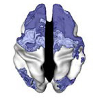
adhd-brain2.jpgEd Yong at Not Exactly Rocket Science reviews new research showing that the brains of children with attention deficit hyperactivity disorder (ADHD) exhibit delayed maturation of the prefrontal cortex and accelerated maturation of the primary motor cortex. The findings are encouraging because they suggest that the brains of patients with ADHD eventually catch with those of nonaffected individuals.

The Neurocritic has a great two part takedown of the New York Times’ latest foray into pseudoscience. He links to several rebuttals and contributes a healthy dose of common sense with this rhetorical question: “Did we really need fMRI to tell us that Mrs. Clinton should try to soften the negative responses of swing voters?”

Language

baboon meta 2

Michael at Shared Symbolic Storage offers a three part review of Baboon Metaphysics, a new book by Dorothy Cheny and Robert Seyfarth. The authors propose that social cognition preceded and made possible the emergence of language, instead of vice versa. Michael argues that this proposal neglects the mechanisms though which language can in turn influence our thought.

“Mama,” “Cookie,” “Nap.” Why do children learn nouns like these before other parts of speech? Dave Munger at Cognitive Daily reports on a creative study in which infants learning their first language were compared to older children learning a second language. This allowed researchers to figure out whether the early abundance of nouns is a function of age or an necessary feature of language acquisition.

That wraps it up for this issue of Encephalon. Bora at A Blog Around the Clock hosts Encephelon #37 on December 3rd. Email your posts to encephalon{dot}host{at}gmail{dot}com or submit using the online form.

Bloggers Against Un-Reviewed Research

I cannot adequately express how pleased I am with the tremendous online outcry against Marco Iacaboni et al.’s recent Op-Ed in the New York Times. After scanning swing voters with fMRI, the authors of this article attempt to draw all sorts of grandious conclusions about what they think of the various presidential candidates.

For instance, the article concludes that “Voters sense both peril and promise in party brands,” and “Emotions about Hillary Clinton are mixed.” Wow, tell me something I don’t know. When the authors get a little more concrete about which emotions are in play, it becomes clear just how speculative their arguments are. When voters viewed Hillary and exhibited activity of the anteriror cingulate, the authors claim that this means they “were battling unacknowledged impulses to like Mrs. Clinton.” However as Daniel Engber points out at Slate, activity of that region could mean many things:

But their interpretation of the Hillary data starts to look a little fishy if you take into account a similar round of FKF brain scans from the last presidential election. In 2004, the same researchers put 20 highly partisan voters into an MRI machine and showed them pictures of George W. Bush, John Kerry, and Ralph Nader. The result: Voters showed heightened activity in the conflict areas—including the anterior cingulate cortex—when they viewed the candidate they hated, as opposed to one they loved. In other words, when a hard-core Democrat looks at a picture of the dreaded George Bush, you get the same brain activity as when a swing voter looks at Hillary Clinton. Suddenly, the Hillary results don’t seem so promising.

While peer review is an imperfect system, it does a good job of preventing scientists from overextending their data to reach speculative conclusions. Reviewers insure that conclusions are based off comparisons of experimental data with control data, something that was definitely lacking from Iacoboni’s research. Because this research was not subjected to these rigorous standards and instead went directly onto the pages of the Times, it is not surprising that the quality of the science is poor.

It’s a shame that respected a neuroscientist like Iacaboni would sell out like this, especially when some of his research is so compelling. However, the debacle does highlight the necessity of peer review. Furthermore, it demonstrates that popular news outlets must learn to cooperate and coexist with scientific journals if they are to succeed at informing readers accurately. When the Times tries to operate outside of the existing peer-review structure, it ends up looking foolish.

For these reasons and more, I support what’s going on over at Bloggers for Peer-Reviewed Research Reporting. But journalistic failures like this one make me wonder if we don’t need another icon as well. So, how about this?

baur2.png

Making Sense of fMRI

Blogging on Peer-Reviewed ResearchFunctional Magnetic Resonance Imaging (fMRI) studies have rapidly become the bread and butter of cognitive neuroscience research. They are equally popular with science journalists who, with the zeal of phrenologists past, gleefully report that “Optimism Lights Up a Brain Part” or “Brain Circuits That Control Hunger Identified.” The only problem with these studies is that nobody knows what the fMRI signal actually means.

Well, that’s an exaggeration. We do know that fMRI signal reflects regional fluctuations in blood oxygenation, but we have yet to figure out what this metabolic activity means in terms of neural activity. In a recent Nature Neuroscience paper, Ahalya Viswanathan and Ralph Freeman make substantial progress toward answering this question.

Increased oxygen consumption might reflect neural inputs (e.g. synaptic activity at dendrites) or it might reflect neural outputs (e.g. action potentials at axons). Viswanathan et al. used a creative experimental design to distinguish between these possibilities. They used dual microelectrodes to simultaneously measure local oxygen levels, spiking activity and local EEG signal (or field potentials) in the cat visual cortex. They found that oxygen levels correlated most closely with the local EEG signal, which is known to reflect dendritic inputs rather than axonal outputs. For a review of this connection please check out my Method of the Month feature.

One challenge in determining whether tissue oxygenation reflects dendritic or axonal activity is that they usually go hand in hand. If a neuron is receiving a flurry of synaptic inputs, it usually starts firing action potentials as well. Viswanathan et al. sidestepped this problem by taking advantage of the fact that the visual system is composed of modules that are arranged in series. Moreover, these modules respond differently to the same visual stimuli. In this study, the experimenters used a drifting pattern of black and white lines to stimulate the visual system. When the grating drifts slowly (e.g. 4 Hz), the visual stimulus activates the thalamus, which goes on to activate the visual cortex. When the grating drifts quickly (e.g. 30 Hz), the stimulus activates the thalamus, but thalamic projections fail to activate the visual cortex. While the cortex receives inputs corresponding to the high frequency visual stimulus, it does not fire action potentials (or spikes) in response.

By recording from the visual cortex, Viswanathan et al. were able to show that both frequencies of visual stimuli resulted in local field potentials and changes in tissue oxygenation, even though only the low fequency stimuli resulted spiking activity. This dissociation strongly suggests that fMRI signals, like EEG signals, reflect synaptic inputs rather than axonal outputs.

Here is one of the most important figures from the paper, which pretty much sums up their findings:

fmri-signal.jpg

On the left, the dark blue histograms show spiking activity in response to visual stimulus (shown by square wave at top) while the waveform shows fluctuations in tissue oxygenation. Note the initial dip in oxygenation followed by a subsequent increase, a response pattern that is typical of regional fMRI signals. On the right, the red areas correspond to increased EEG signal upon stimulus presentation. The 30 Hz signal alters blood oxygenation and local field potentials without resulting in action potentials.

This study clarifies one of the major questions obscuring the accurate interpretation of fMRI data. Neural inputs turn out to matter more than neural outputs. Unfortunately, it doesn’t clear up the whole mystery surrounding imaging data. For instance, if the cingulate cortex lights up during optimistic thoughts, we still don’t know the source of those synaptic inputs. Most brain regions integrate inputs from disparate brain regions, and it looks like the fMRI signal won’t be able to distinguish among these.

Nonetheless, these studies are a step in the right direction: away from neo-phrenology and toward a functional understanding of fMRI data.

Reference:

Viswanathan, A. and Freeman, R.D. Neurometabolic coupling in cerebral cortex reflects synaptic more than spiking activity. Nat. Neurosci. 10, 1308-12 (2007)

I Was a Subject in Deena Weisberg’s Study

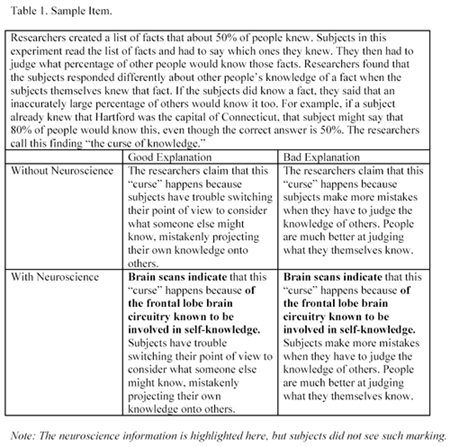
Two years ago I took a cognitive neuroscience course in which all the students were asked to participate in a psychological study. We filled out an online survey that asked us to rate various explanations for psychological phenomena. I remember hating the survey because all the explanations seemed bad, because it was time-consuming, and because I had no idea what the study was about. Needless to say, I treated it with a healthy dose of apathy.

Now the study, led by PhD student Deena Weisberg, is available online in advance of its publication in the Journal of Cognitive Neuroscience. Apparently it was meant to test how irrelevant neuroscience information can bias our judgments about whether an explanation is good or bad. Here is an example survey question:

weisberg et al 3

Weisberg asked novices, neuroscience students, and experts to take the survey. She found that extraneous neuroscience information made novices and students more likely to endorse bad explanations. Experts did not fall for the neuro-jargon. In fact, they were less likely to endorse the good explanations if they included extraneous information.

Weisberg concludes that we should be careful with our appeals to neuroscience information, especially when the audience is composed of non-experts. For instance, if scientific evidence is presented in a courtroom, jurors might allow it to sway their judgment even if it is irrelevant.

So how does it feel being held up to the scientific community as an exemplar idiot? Well, it’s a bit embarrassing. One of my coping mechanisms has been to criticize the experimental design. For instance, I think its problematic that the with neuroscience explanations were longer than the without neuroscience explantions. If subjects merely skimmed some of the questions (not that I would ever do such a thing), they might be more likely to endorse lengthier explanations.

Another problem I have is the circularity of the rating system. The ‘objective’ ratings for whether explanations were good or bad, and for whether the extra information was indeed irrelevant, were supplied by cognitive neuroscience experts. Is it really surprising then, that this group would show the least amount of bias in judging the explanations?

I could rattle off a few more complaints, but I’ll stop here. In fact, I think the study deserves a lot of the attention that it is getting. With new fMRI studies coming out daily, it’s important not to get sucked in by all the pretty pictures and retain some critical distance.

Neurons for Counting

A group of Duke University scientists have just published an interesting study in the open-access PLoS Biology Journal. They wanted to verify the existence of so-called integrator neurons whose activity reflects the number of items in their receptive field. To get monkeys to pay attention to the quantities, Roitman et al. used a paradigm in which monkeys received a reward for delayed eye movement toward a previously displayed target. During this task, the experimenters also presented the monkeys with a group of dots that represented the size of the reward that they would receive upon successful task completion. The basic design is illustrated below:

Roitman et al. simultaneously measured the activity of single neurons in the monkey’s parietal lobe. They found one subset of neurons in the lateral intraparietal area (LIP) that activated more strongly to larger numbers than to smaller numbers. Moreover, they found another group of neurons in the LIP that activated more strongly to smaller numbers than to larger numbers. Both groups of neurons exhibited an initial increase in activity upon presentation of the number cue, but differences were found in how fast the activity decayed. The exact timecourse can be seen below:

Roitman et al. hypothesize that neurons exhibiting a graded response to numerical quantity provide crucial inputs for cells tuned to cardinal numbers (these cells have already been located in the prefrontal cortex and parietal lobe). In other words the parietal cortex would first estimate quantity and then translate this estimate into an integral value. Alternatively, it is possible that the information flows the opposite direction and general estimates of quantity follow more discrete identification of cardinal numbers. Hopefully future studies will be able to discriminate between these hypotheses.